Navicular Disease
One of the major diseases in horses, especially in the warmblood horses as well as in the American Quarterhorse, navicular disease is a common problem, limiting an athletic career of the horse.
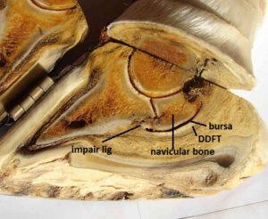
Recent research has shown that not only the navicular bone, in the past thought to be the major component in navicular disease, is an important factor. We know now that the Deep Digital Flexor Tendon, the Impair ligament and the collateral ligaments of the navicular bone and the navicular bursa are anatomical structures that are involved in a large part of the cases with navicular disease.
 It seems now that most navicular disease cases start with an injury to the Deep Digital Flexor Tendon (see the diagram left). Where this tendon makes an angle to pass the navicular bone, this is a region where the tendon can be damaged. Fibrils of the tendon will be pulled of the core and float free in the bursa, the liquid filled bag in between the tendon and the navicular bone. These fibrils will irritate the bursa, making it to produce more fluid (synovia) and symptoms of inflammation will occur (more fluid, higher pressure in the bursa, pain, etc) After a while the cartilage at the flexor side of the navicular bone will get damaged. When the damage to the cartilage is more severe, also the bone underneath it will start to change (more dense, more bone, this will be visible on an X-ray and is called sclerosis) and the synovial membrane (the inside layer of the bursa that is producing the fluid) will proliferate and finally invade the navicular bone (giving the bone on X-ray the specific appearance with thewidened and lollypop-shaped channels.
This is more or less the end stage of navicular disease, and in this stage not a lot of options to fix this are available.
 In earlier stages however, when there is no damage to the navicular bone ( and the X-rays will show a normal appaerance) there are several options for treatment available.
With radiography (X-rays) we can obtain nice images of the navicular bone, especially when we use different viewing angles (lateral and anterior-posterior images, skyline view) and the use of digital X-ray equipment. But only the bony structures can be evaluated with X-rays.
Information about the navicular bursa, the Deep Digital Flexor Tendon and the Impair ligament and collateral ligaments, as well as the cartilage on the flexor side of the navicular bone is not accessible with X-rays.
MRI can give an enormous amount of information about these soft tissue structures, but it needs general anesthesia. Because of that it is quite expensive.
When over time follow up is needed, MRI is becoming very expensive (nearly nobody will accept an MRI exam every 2 months for ½ to 1 year)
For that reason de doctors at the Equine All-Sports Medicine Center have developed an ultrasound technique that can visualize the DDFT, the bursa and the cartilage of the navicular bone, and sometimes even the impair ligament and the collateral ligaments.
They have published about that technique in the veterinary textbook “Current Therapy in veterinary Medicine” Ed 6, N.E Robinson and K Sprayberry, editors. Chapter 124: Ultrasound of the distal limb by Dr R van Wessum.
Dr Johnston has developed in cooperation with Dr Nickels of MSU a surgical technique to debride the DDFT and the Bursa when scar tissue or inflammation is present.
 The EASMC is one of the few facilities in the world that offers the options to evaluate the navicular region with ultrasound and has the opportunity to perform arthroscopic surgery to the navicular bursa (called bursoscopy), a minimal invasive technique, as a top-notch treatment modality for several navicular disease cases.
Especially the surgical treatment option is top notch and developed very recently, and it is the best available treatment modality when the DDF tendon is damaged and has material in the bursa. Not any other treatment will remove this material, so not any other technique will reduce inflammation and stimulate healing. All other treatment options just deal with symptoms.
Other treatment options are available however, and they are good choices when surgery is not an option.
Corrective and orthopedic shoeing can reduce the problems, caused by navicular disease. Very often we will advise a shoe with a slight wedged hoofpad and a breakover point that is trimmed back, so forces on the navicular region are reduced.
With our in-house farrier we can achieve the best possible adaptation for your horse’s feet in case of navicular disease.
Shoeing is quite often combined with medication.
Most common medical treatment of navicular disease is the use of NSAIDs, substances like Aspirin, phenylbutazone and firocoxib (Equioxx). They will reduce inflammation and pain, but are not really treating the problem, just make it more manageable.
Injections of medication (mostly corticosteroids) into the coffin joint or into the bursa can assist in reducing inflammation in the navicular region. Especially the injection of the navicular bursa is not very easy, and we use ultrasound guidance or radiographic guidance to know for sure we are injecting the bursa.
A treatment option that is not FDA-approved in the US, but has been available in Europe for some years is the use of Tildren. This medication will reduce the resorption of bone (when the vascular channels in the navicular bone are enlarged and visible on X-rays) and so enhance healing and limit pain.
At the EASMC we can assist you in making a good evaluation of the navicular region and come to a funded and scientifically based advise for treatment and management, so your horse can perform better.